Overview
At Pawar Radiology Center, our advanced colour Doppler ultrasound service reveals real‑time blood flow patterns with exceptional clarity. Under the guidance of Dr Harish Pawar, your scans are interpreted by the Best Radiologist in Thane, delivering accurate insights to support your care.
What Is Colour Doppler Sonography?
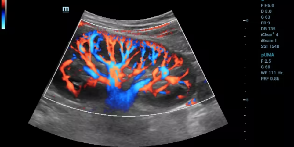
Ultrasound colour Doppler, often called Doppler, uses sound waves to visualize blood movement within vessels. This non‑invasive examination helps identify blockages, assess vessel health, and guide treatment plans by overlaying flow data in vivid colours. Unlike traditional ultrasound, Doppler highlights velocity and direction, making it invaluable for vascular assessments.
Why Choose Pawar Radiology Center?
Expertise & E‑E‑A‑T Focus
- Dr. Harish Pawar brings over 15 years of specialized training in vascular imaging. His depth of Experience, Expertise, Authoritativeness, and Trustworthiness ensure your Doppler study is interpreted precisely.
State‑of‑the‑Art Equipment
- We employ high‑resolution probes and the latest software algorithms, guaranteeing you receive the clearest possible colour Doppler sonography images.
Patient‑Centered Comfort
- From private exam suites to empathetic sonographers, your comfort and privacy are top priorities.
Rapid Reporting
- Preliminary findings are shared immediately after your scan, with a comprehensive digital report—complete with annotated color stills—delivered within 24 hours.
Our Colour Doppler Sonography Services
We offer specialized Doppler studies tailored to four critical areas:
- Limbs Doppler
- Portal Doppler
- Renal Doppler
- Scrotal Doppler
Each service uses the same advanced principles of colour doppler sonography near me, adapted to that region's unique anatomy and clinical questions.
1. Limb Doppler Ultrasound
Purpose & Benefits
A limb Doppler ultrasound evaluates arterial and venous circulation in the arms or legs. It can detect:
- Deep vein thrombosis (DVT): Clots that pose a risk of pulmonary embolism.
- Peripheral arterial disease (PAD): Narrowing or blockages that cause leg pain or wounds that won’t heal.
- Chronic venous insufficiency: Valve dysfunction leading to varicose veins and swelling.
How It Works
A warm gel is applied to your limb, and a linear transducer sends color‑coded echoes back to the machine, mapping flow speed and direction. A limb Doppler sonography exam typically takes 20–30 minutes.
When to Book
Seek a limb doppler ultrasound near me if you experience leg cramps, persistent swelling, or visible veins that bulge and ache. Early detection can prevent serious complications.
2. Portal Vein Doppler Ultrasound
Purpose & Benefits
A portal vein doppler ultrasound examines blood flow through the liver’s portal vein system. This test is crucial for:
- Cirrhosis monitoring: Assess elevated liver pressures (portal hypertension).
- Pre‑ and post‑transplant evaluation: Ensure proper perfusion in donor and recipient.
- Detecting thrombosis: Identify clots in the portal vein that can lead to liver failure.
Procedure Details
After fasting for 6 hours, you lie on your back. A curved‑array probe scans beneath your ribcage. Color maps reveal flow velocity and direction—key data for portal doppler ultrasound assessments.
Clinical Impact
Timely portal vein doppler radiology informs interventions like shunt placement or anticoagulation therapy, improving patient outcomes in chronic liver disease.
3. Renal Doppler Ultrasound
Purpose & Benefits
A Renal doppler sonography evaluates blood flow to and within the kidneys. It aids in diagnosing:
- Renovascular hypertension: High blood pressure caused by artery narrowing.
- Renal artery stenosis: Blockages that can lead to kidney damage.
- Transplant assessment: Monitor blood flow in transplanted kidneys.
Exam Process
You’ll lie on your back with a pillow under one side. A curved probe scans each kidney in multiple planes. The color overlay highlights any flow irregularities, guiding further tests or treatments.
Why It Matters
Early detection of renal vascular issues prevents irreversible kidney damage and helps manage resistant hypertension effectively.
4. Scrotal Doppler Ultrasound
Purpose & Benefits
A scrotal doppler scan examines blood flow within the testicles and surrounding structures. It identifies:
- Varicocele: Enlarged veins that can impair fertility.
- Testicular torsion: A surgical emergency where blood flow is cut off.
- Inflammation or infection: Epididymitis or orchitis.
- Tumor vascularity: Characterize masses based on blood supply.
Procedure Details
You’ll lie with a towel draped for discretion. A generous gel coat and a small linear probe reveal live flow maps. A typical usg Scrotal doppler takes 15–20 minutes.
Key Takeaway
Rapid, accurate detection of testicular torsion can save testicular function. Evaluating scrotal Doppler ultrasound early ensures prompt surgical or medical treatment.
Preparing for Your Colour Doppler Sonography
To ensure optimal image quality and a smooth appointment:
Fasting Requirements
For portal vein doppler ultrasound, avoid food and drink (except water) 6–8 hours prior.
Comfortable Clothing
Wear loose garments that allow easy access to the scan area—especially for limb or scrotal studies.
Medical History
Bring any relevant imaging reports, surgical histories, or lab results to help Dr. Pawar interpret flow patterns in context.
Our friendly scheduling team will send a personalized reminder, including specifics for each Doppler study, and information about colour doppler sonography price.
What to Expect During Your Appointment
Warm Welcome & Check‑In
Confirm your details, referrals, and discuss any questions about colour doppler sonography price or insurance coverage.
Private Exam Room
You’ll change in privacy. The sonographer will explain the procedure and position you comfortably.
Gel & Probe Application
A water‑based gel eases probe movement and enhances sound‑wave transmission.
Real‑Time Imaging
Watch color flow maps on a high‑resolution monitor as the sonographer captures multiple views.
Immediate Feedback
Dr. Pawar reviews key images, discusses preliminary findings, and takes additional views if needed.
Digital Report Delivery
Receive a detailed, annotated report within 24 hours—access it securely online or via our patient portal.
Take the Next Step in Vascular Imaging
Don’t let circulatory issues go undetected. Trust the Best Radiologist in Thane for your colour Doppler sonography needs. Whether you require a limb Doppler ultrasound, portal vein doppler radiology, Renal doppler ultrasound near me, or Scrotal doppler scan, our center combines cutting‑edge technology with expert interpretation. Book today to gain the clarity you need for confident, timely care.
Call us at +91-9967677602 or Schedule Online to reserve your appointment. Your vascular health is our priority.
Frequently Asked Questions
1. How do I book a “colour doppler sonography near me”?
Ans : Pawar Radiology Center serves Thane and Hiranandani Estate. Call +91-9967677602 or book online to schedule your Doppler study.
2. Will I know the “colour doppler sonography price” beforehand?
Ans : Yes. Our staff provides transparent cost estimates during booking, including any add‑ons for advanced 3D imaging or guided biopsies.
3.Are Doppler ultrasounds safe?
Ans : Absolutely. Doppler uses harmless sound waves—there’s no radiation, making it safe even for pregnant women when evaluating limb circulation or renal flow.
4. How long does each Doppler exam take?
Ans : Most studies finish within 15–30 minutes, depending on complexity and patient comfort.
5. When will I get my results?
Ans : Preliminary results are shared immediately post‑scan. A full digital report, complete with color stills, is ready within 24 hours.